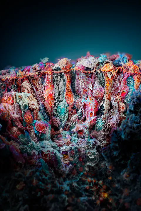
For many years, I was a distant admirer of Martin Oeggerli, who is without doubt one of the most prolific science photographers alive, an artist who captures the molecular and cellular world with such audacity and clarity that every image feels like a doorway into a new universe. When I saw one of his photographs for the first time a decade ago, I could hardly understand how it had been created. My wonder was so great that I never even considered contacting him. But chance intervened when we finally crossed paths at the Novartis Pavillon in 2022. A brief conversation turned into a long one, and soon afterward we began a collaboration that has now stretched across several covers for Live Magazine and led to the development of the Novartis science photography. Through that journey I discovered not only how Oeggerli works, where he draws inspiration, and what drives him artistically, but also how deeply he feels the responsibility of translating the hidden world into something the human eye and heart can grasp.
Martin Oeggerli in a lab in Basel. As he does not own a high-resolution microscope himself, he often works in different research locations in such as the C-CINA at the Biozentrum of the University of Basel, the University Hospital of Basel (Department of Pathology), and the University of Applied Sciences Northwestern Switzerland (FHNW) in Muttenz.
Following him into the laboratory at the University of Basel, observing the way he developed our mast cell and Ferrari-red covers and later the unique Novartis science images, watching his patient, meticulous approach to the scanning electron microscope and the extraordinary care he brings to the colorization of each image, left me speechless. If anything, it deepened the sense of wonder I felt the first time I encountered one of his magical windows into the microscopic universe beneath our feet and under our skin.
Pollen of the foxglove flower.
Coolpix explosion To understand how Martin Oeggerli became one of the world’s most distinctive scientific photographers, it helps to begin in the neighborhood where he grew up. “I had a very nice childhood, close to a forest,” he told me during our interview that took place at the end of 2025. “I played a lot there. I was always very interested in animals and plants.” His curiosity about nature – exemplified in his early color drawings – was present long before he ever held a camera, but photography entered his life in a way that feels almost cinematic. His father, an amateur photographer, recognized Martin’s fascination and quietly nurtured it. “One day I came home and found a big package in front of my door,” he said. “A Coolpix camera. A gift from my parents out of the blue.” He still laughs at the memory of what happened next. “I think I did around 20,000 pictures in the first month,” he said. “All brakes were off.” With digital photography he discovered the addictive immediacy of seeing results instantly, refining them, learning through repetition. It was the beginning of a relationship between observation and expression that would shape his life.
At first, the world remained at human scale. He took pictures of reptile exhibitions, flowers in the botanical garden, or the strange and beautiful structures of insects. But he soon felt the pull toward smaller realms. “I realized there is this world you cannot see with a standard camera,” he said. “And with a scanning electron microscope, you are in the middle of it.” The leap into that world would come through an unexpected door.
SEM images While finishing his Ph.D., Oeggerli earned additional income working as a tennis instructor for a chemical company in Basel. One day they approached him with an unusual request: Do you know anyone with a scanning electron microscope? They needed SEM images for their annual report. “We had just one month left,” he recalled. “Those were my first 14 images.” Without formal training in electron microscopy, he embarked on a crash course in a field that demands both scientific rigor and artistic sensitivity. More remarkably, he also taught himself to colorize the images, an unheard-of leap for a newcomer. “I taught myself how to color them in that very short time,” he said. “Later I developed my own technique, different from what graphic students learn. It gave me an advantage; I could use it more creatively.” When he finished the project and went on holiday to New Zealand, he returned with a powerful sense of direction. “I came back and had this unsatiable hunger,” he said. “I wanted to go for even more attractive things I knew existed.” What followed was the first body of work that would carry his name far beyond Basel: a long, solitary exploration of pollen grains. Breakthrough recognition For one and a half years, Martin collected, prepared, imaged, and colorized pollen grains, those tiny, often unnoticed specks that become colossal worlds under the electron microscope. When he finally gathered the courage to send them to National Geographic, the response he initially received was anything but encouraging. “They told me: We only work with the best photographers in the world, and I do not know your name,” Martin said. “You can send something, but don’t be disappointed if you never hear anything again.” He sent them anyway. A year passed. Silence. And then, suddenly, a message: As you probably already know, your article has been accepted. Martin still shakes his head at the memory. “I thought they must have sent it to the wrong person,” he said. “I expected an e-mail three days later saying, sorry, that was not for you.”
But it was real. And when he sent them early images of butterfly eggs, the answer came in just three words: “Beautiful, beautiful, beautiful.” It was a moment that transformed his path. “That was when my hobby became a profession,” he said. From that point on, Martin began to build a new visual language for the invisible world. Envisioning the invisible The defining element of Oeggerli’s work – who also publishes under his nom de plume Micronaut – is his use of color. This is not decorative color, nor arbitrary enhancement. For Oeggerli, color is both a scientific translation and an artistic philosophy. “The only thing you don’t have with the electron microscope is color,” he said. “By coloring these grayscale images, you can revive them. You can start to tell a story.” He aims for a photorealistic impression: “I don’t want someone to immediately realize that it’s artificially colored. If they think it’s a camera photograph at first glance, I’ve done my job.” Achieving that impression takes a lot of time. “I invest so much time into a single picture,” he said. “I am only capable of capturing maybe 12 to 20 images a year.”
But what he produces are not simply images, they are portals. And the way he constructs them is a kind of chromatic alchemy. Alchemy In the world of scanning electron microscopy, there is no color, only the stark terrain of grayscale. What Oeggerli does is nothing less than invent a color universe for structures that have never been seen in light. And he does it with extraordinary discipline. “When you look at a tree, you think you see green,” he said. “But if you look closely, there are shades of yellow, red, different greens. I mix subtly different colors to create a photorealistic impression.” He begins by masking forms, sometimes hundreds of them, sometimes thousands, each one defined by biological shape, function, or surface texture. These masks become the scaffolding of his chromatic world. “With the electron microscope you are lost,” he told me. “You don’t know what you are seeing. Color becomes the only guide you have.”
Timelaspe of coloring
Starting from black-and-white scans, the colorization process is extremely work-intensive. Martin Oeggerli often creates hundreds of layers to create an image. The process can take weeks to months, depending on the structures of a sample.
Color becomes navigation, orientation, and narrative all at once. A mast cell might glow with warm, fiery hues to evoke biological urgency. The basil leaf’s secret glands, which hold the essential stores of aromatic compounds, might shimmer with layered greens and ochres. In a dividing bacterium, the thin membrane separating daughter cells may be rendered in near-white tones to convey fragility. “Color lets me tell a story biology alone cannot show,” he said. “It’s the difference between looking and understanding.” For a Live Magazine issue dedicated to color, Oeggerli’s work becomes almost emblematic: he is not coloring the microscopic world; he is giving it light, depth, emotion, and legibility, a chromatic language for a universe that has never seen color at all. Retina and corals Nowhere is this fusion of scientific insight and aesthetic sensitivity more striking than in Oeggerli’s image of the human retina. “The retina has 140 million receptors,” he told me, his voice shifting into that quiet register usually reserved for awe or reverence. “It can send signals 18 times a second through the optic nerve. It gives me shivers when I think about it.”
The artificial retina was created at the Institute of Molecular and Clinical Ophthalmology Basel (IOB) and served as the cover for live #19.
Colorizing the retina image was an undertaking that required both patience and reverence. “There were so many fibers,” he recalled. “I colored them individually just to make them visible.” The result is not merely a depiction of anatomy; it is a portrait of vision itself, a stained-glass window of biological engineering. Not every image Oeggerli creates comes from meticulous planning. Some come from surprise, serendipity, or sheer scientific curiosity. One of his most famous images came from human microbiome material. “As a biologist, I should have known how many bacteria there are in the gut,” he admitted. “But I was not expecting a picture that looked like a coral reef.” The result is a landscape that feels oceanic, architectural, and alien all at once. It is a reminder that complexity does not diminish with scale; it becomes more astonishing.
Perhaps the most surreal work in Oeggerli’s portfolio is the surface of a Chilean stinging nettle, a botanical rarity grown in a locked greenhouse at the Basel botanical garden. The microscopic view is astonishing: glands on stalks, crystalline formations, burning hairs jutting out like defensive architecture. “It looks surreal,” Oeggerli said. “But it is the surface of a plant.” More to come Although Oeggerli approaches microscopy with an artist’s sensibility, his images have profound meaning for scientists too. “Often they work with PCR or other methods and never see what they research,” he explained. “But with a picture, suddenly they can.” He once showed a mycologist who had spent 30 years studying fungi a SEM image of fungal spores from a plant growing on Oeggerli’s balcony. “For the first time, he really saw the spores he’d been researching,” Oeggerli said. “He couldn’t believe his eyes.” This is the power of Oeggerli’s art: it returns the invisible to the realm of the visible, restoring an emotional connection between scientists and their subjects. Among Oeggerli’s most magnified images is a dividing Streptococcus bacterium. “When printed large,” he said, “it is more than a million times magnified.” And yet what strikes him most is not the scale but the delicacy. “I colored the dividing part in a very light tone,” he said. “I wanted to indicate the thinness of the skin between the daughter cells.” Here again, color becomes meaning, an informative and emotional device. When asked whether he sees limits or a unifying theme in his work, Oeggerli answers simply: “Curiosity. That is the first thing.” He is aware that below bacteria lie viruses, and beneath viruses lie realms beyond the reach of the electron microscope. But rather than seeing boundaries, he sees infinite beginnings.
“There is no end,” he said. “If I find something beautiful, I dig deeper. I look for related species. I look for new structures. There is always more.” Oeggerli’s world is inexhaustible. And that is the point. “We think we know the world, but maybe we haven’t seen 99 percent of it.” Whether exploring pollen grains, basil leaves, mast cells, fungal spores, or bacteria dividing into daughter cells, Martin Oeggerli achieves something rare: He not only documents the microscopic world, he also translates it into human experience. “There are so many things we never see,” he told me. “But I try to show them, so that someone joining me on this expedition into the invisible universe can feel oriented, can understand, can be touched.”
One new element in the Novartis brand image library is this immune cell by Martin Oggerli.
Thanks to Oeggerli’s unique way of portraying the world, Novartis’s scientific work can now also be viewed in a fresh and unconventional way and be experienced in a new light, offering scientists, patients and the public at large a new perspective on the company’s work.
Martin Oeggerli working at the Novartis Pavillon during the 2026 summer exhibition that focused on some of his most recent works.
People Taking a closer look. Translating the invisible Text Goran Mijuk. Photos Sophia Lavater and Martin Oeggerli. Videos Goran Mijuk. Martin Oeggerli’s way into science photography was the result of his deep fascination with nature from an early age coupled with a random call for an annual report feature. In the span of a few years, the Swiss native won world-renown for his unique micro photography, which will now also be part of Novartis’ brand identity.
The image-making process.
Before starting to work on the image, sample preparation is key. Besides drying the sample with alcohol, the samples are coated with gold. Oeggerl needs to work with magnifying glasses.
The samples are put on round chips to fit into the scanning microscopes.
The samples are gold-plated to ensure electrical conductivity, prevent charging artifacts, and enhance secondary electron emission for high-resolution SEM imaging.
Close-up of gold-plated samples.
Scanning electron microscopes work differently from regular microscopes. They use a focused beam of electrons rather than visible light to examine the surface of specimens at extremely high magnification.
After sample preparation and upload on the SEM, the actual work begins. Martin Oeggerli navigates the sample for the most fitting subject. This process can take hours and even days. Often, the samples may not yield what he wishes to later depict in his images.
You may also be interested in the following:

Thanks for reading Live.Magazine
Stay connected by subscribing to our newsletter
SubscribeBy submitting your email, you consent to Novartis AG collecting and processing your email data for Novartis internal use, in accordance with our privacy policy, and by protected technical means.